Malignant Mesothelioma Ct Radiology - Metastatic Biphasic Pleural Mesothelioma Presenting With Cauda Equina Syndrome Sciencedirect : malignant pleural mesothelioma is a rare tumor.. mesothelioma has a long latency period of 20 to 40 years, and many patients do not have symptoms until the disease is in its later stages, when metastasis is more likely to occur. Staging of malignant pleural mesothelioma: There is a large pleural effusion present. Ajr am j roentgenol 1981; malignant mesothelioma and metastatic adenocarcinoma are the two most common pleural malignancies.
They have similar imaging findings but different prognosis and treatment. ct manifestations in 50 cases. Role of ct, mri, and pet/ct in staging evaluation and treatment considerations. In each case the ct showed. mesothelioma is a malignant tumor that arises from the mesothelial surfaces of the pleural and peritoneal cavities, the tunica vaginalis, or the pericardium.
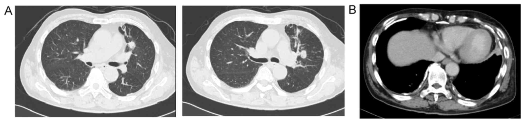
Double Cancer Comprising Malignant Pleural Mesothelioma And Squamous Cell Carcinoma Of The Lung Treated With Radiotherapy A Case Report from www.spandidos-publications.com malignant mesothelioma and metastatic adenocarcinoma are the two most common pleural malignancies. A) axial ct scan, nodular pleural thickening (white arrow), b) sagittal chest ct scan of the soft tissue. Computed tomography is the primary imaging method used for the diagnosis and the staging of malignant mesothelioma, but also for guiding biopsy for tissue diagnosis. This is a case of pathology proven metastatic malignant mesothelioma. Leave a reply cancel reply. The radiology of thoracic malignant mesothelioma. They have similar imaging findings but different prognosis and treatment. There are few reports about ct and mri findings about malignant mesothelioma of the tunica vaginalis testis.
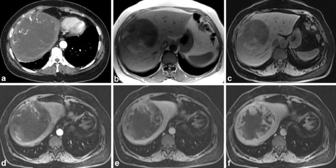
Magnetic resonans imaging (mri) is useful for detection of extension of disease, especially to the chest wall and diaphragm (moore et al., 2008, wang et al., 2004).
ct of malignant pleural mesothelioma. Leave a reply cancel reply. Although the chest film findings of pleural mesothelioma are well described, there are few descriptions of the findings of computed tomography (ct). 60 year old male with left sided chest pain. Computed tomography (ct) enables early detection of small pleural tumors and pleural effusions and definition of the extension of the tumor along the pleural surfaces and fissures. Computed tomography is the primary imaging method used for the diagnosis and the staging of malignant mesothelioma, but also for guiding biopsy for tissue diagnosis. Doctors can make a metastatic mesothelioma diagnosis through radiology imaging scans such as mri, ct or pet. malignant peritoneal mesothelioma is an unusual disease process characterized radiologically by ascites and infiltration of the peritoneum by multiple small tumor nodules.both parietal and visceral peritoneum are involved by the multiple malignant tumor nodules. mesothelioma lawsuit radiology mesothelioma radiology imaging from i.pinimg.com. Kitajima k, doi h, kuribayashi k, et al. Staging of malignant pleural mesothelioma: Methods the patient cohort included nine patients undergoing chemotherapy and five patients on observation. Superior vena cava syndrome with malignant reasons nejm.
In 2016, mesothelioma of any site had an incidence rate of 2.0 cases per 100,000 men and 0.3 per 100,000 women in italy (), with pleural localization as the commonest site (93% of cases) ().however, in some geographic areas, incidence has considerably increased over the last decades due to professional exposure to. University of michigan medical school, ann arbor, mi 48105. Sheets of tumor can be followed inferiorly along the diaphragmatic crura. In only one patient was. Symptoms or abnormalities not consistent with the initial.
An Unusual Liver Mass Primary Malignant Mesothelioma Of The Liver Springerlink from media.springernature.com malignant mesothelioma early detection, diagnosis, and staging cancer.org | 1.800.227.2345 detection and diagnosis finding cancer early, when it's small and before it has spread, often allows for more Coolen j, de keyzer f, nafteux p, et al. 60 year old male with left sided chest pain. Initial ultrasound examination demonstrated the patient's known infrarenal abdominal aortic aneurysm and discovered a 3 × 4 cm solid, slightly lobulated, hypoechoic mass contiguous with the upper pole of the left kidney and adjacent to the spleen. Kitajima k, doi h, kuribayashi k, et al. Peritoneal cancer index (pci), as assessed by ct, is utiliz … Sixty percent occur in the right pleural cavity and 40% in the left. Ajr am j roentgenol 1981;
malignant mesothelioma and metastatic adenocarcinoma are the two most common pleural malignancies.
mesothelioma lawsuit radiology mesothelioma radiology imaging from i.pinimg.com. malignant pleural mesothelioma is a rare tumor. Staging of malignant pleural mesothelioma: Kitajima k, doi h, kuribayashi k, et al. Multidetector ct findings and differential diagnoses of malignant pleural mesothelioma and metastatic pleural diseases in korea. malignant pleural mesothelioma (mpm) is an uncommon neoplasia. Computed tomography (ct) enables early detection of small pleural tumors and pleural effusions and definition of the extension of the tumor along the pleural surfaces and fissures. Imaging plays an essential role in the evaluation of malignant pleural mesothelioma (mpm). A cancer diagnosis may be overwhelming. Doctors can make a metastatic mesothelioma diagnosis through radiology imaging scans such as mri, ct or pet. Initial ultrasound examination demonstrated the patient's known infrarenal abdominal aortic aneurysm and discovered a 3 × 4 cm solid, slightly lobulated, hypoechoic mass contiguous with the upper pole of the left kidney and adjacent to the spleen. mesothelioma is an uncommon malignant tumor that arises from mesenchymal tissue and involves pleura, peritoneum, pericardium and very rarely, the tunica vaginalis (< Asbestos plaques are not a…
For the evaluation of lymph nodes, internal mammary nodes were considered abnormal when Sheets of tumor can be followed inferiorly along the diaphragmatic crura. malignant pleural mesothelioma is a rare tumor. This is an uncommon benign primary peritoneal tumor that has no relation with the malignant mesothelioma. Oncology case 7 june 19, 2019;
Malignant Pleural Mesothelioma Radiology Case Radiopaedia Org from prod-images-static.radiopaedia.org ct of malignant pleural mesothelioma. Your email address will not be published. The second most common radiologic finding was an infiltration of the small bowel mesentery by tumor reported in 41% of patients 2 . Leung an, müller nl, miller rr. Symptoms or abnormalities not consistent with the initial. There's no history of syncope or. malignant pleural mesothelioma is a rare tumor. University of michigan medical school, ann arbor, mi 48105.
Methods the patient cohort included nine patients undergoing chemotherapy and five patients on observation.
The radiology of thoracic malignant mesothelioma. Use of ct for tumor staging. Imaging plays an essential role in the diagnosis, staging, and clinical management of patients with mesothelioma. Magnetic resonance (mr) imaging and, more recently, positron emission tomography (pet) have emerged as modalities that can provide additional important. mesothelioma is an uncommon malignant tumor that arises from mesenchymal tissue and involves pleura, peritoneum, pericardium and very rarely, the tunica vaginalis (< Heelan rt, rusch vw, begg cb, et al. There are unusual radiologic presentations of malignant peritoneal mesothelioma that are important to recognize in order to. ct in differential diagnosis of diffuse pleural. This prior report indicated that 43% of patients had ascites 2 . Symptoms or abnormalities not consistent with the initial. Richmond radiology service (114) veterans administration medical center and department of radiology. Ajr am j roentgenol 1981; mesothelioma lawsuit radiology mesothelioma radiology imaging from i.pinimg.com.


0 Comments